As also called a retrusive lower jaw, the lower jaw shifts backward in order to chew food. The lower teeth are positioned deeply behind the upper teeth. In many cases, the improper occlusion is caused by placing inlays and crowns lower than where they should be. Many people are not aware of the condition until it results in other problems, including TMJ, a cracked tooth, cavities or other periodontal diseases. A patient complains of uncomfortable back teeth, but a general dentist does not see any particular cause. We often find the malocclusion as a source of the problem.
Many people have either inlays or crowns. In order to have a correct bite, adjusting the height of the fillings is very important. It is easy to notice if the fillings are too high. However, a patient is often not able to detect a filling that is too low.
The uneven height creates a space between the upper and lower teeth, which causes an inability to chew properly. Chewing habits gradually and unconsciously change. As a result, the lower jaw slowly shifts to backward to chew food.
The retrusive lower jaw stress on the back teeth often causes periodontal diseases, cavities and tooth cracks. It may also result in many other problems, including jaw pain, stiff shoulders, headaches and TMJ.
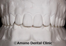
This photograph shows the front teeth of the patient with a retrusive lower jaw. The upper front teeth clearly cover the lower front teeth. All the stress goes on the back teeth.
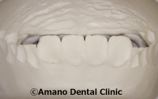
The second photograph shows the front teeth from inside of the mouth. Tips of the lower front teeth bite into the gum, instead of touching the upper front teeth. Cusps (canine teeth) no longer function to tear and grip food, and an improper bite occlusion occurs.
Please click here to read more about a cuspid guidance occlusion.
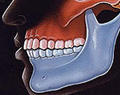
This is a side view. Compared to the normal bite condition shown below, the lower front teeth are located far back. It also shows that the lower back molar tooth is pushed backward than the upper back molar tooth.
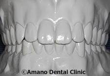
The photograph shows a front view of the proper occlusion. The lower teeth are visible from the front.
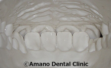
This shows the front teeth from inside of the mouth. In the proper occlusion, the lower teeth touch the inner surface of the upper teeth, and cusps are also in line.
This is a side view of the proper occlusion. It clearly shows that the upper and lower front teeth are in normal position. Upper and lower back teeth also meet properly to maintain abilities to chew food.
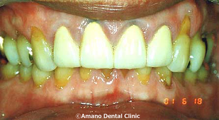
This patient complained about swolling and pain in the back teeth. He also suffered from a severe stiff shoulder and an headache. He went to a chiropractor once a week to ease the pain.
We found that crowns and fillers of the back teeth were so low that he had a deep-bite condition. As shown in the photograph, the lower front teeth are hidden behind the upper front teeth, which is a common sign of malocclusion. Additionally, we found that gums around the front crowns were discolored and swollen due to the ion of the metal crowns.
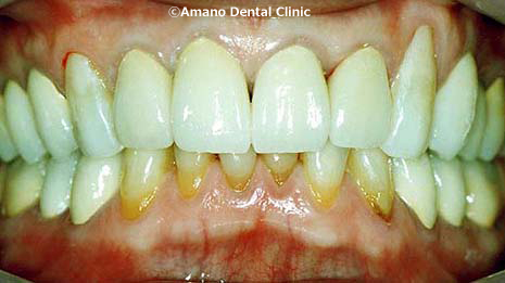
The second photograph shows the front teeth after the treatment. After the basic gum treatment, the metal crowns were replaced withzirconia all-ceramic crowns, which contain no metal. The new crowns were carefully aligned. Also, we created the ideal occlusion (cuspid guidance occlusion) by applying the new crowns.
The condition improved tremendously. The lower front teeth became more visible, and the healthy gum was also restored. Because he no longer suffers from stiff shoulder or headache, he does not need to see a chiropractor.